Breast Cancer Risk Analysis
Mammography Biomarkers for Personalized Screening and Clinical Studies

Mads Nielsen, PhD
The BreastIQ framework from Biomediq offers automatic, computer-based analysis of mammograms for analysis of future risk of breast cancer.
- The package provides:
- Fully automatic segmentation of breast region in film-based and direct digital mammograms.
- Computation of mammographic density.
- Novel texture markers BC MTR of breast cancer risk based on recognition of local patterns of dense tissue.
- Novel texture markers HRT MTR of architectural changes of parenchymal patterns related to various regimes of HRT.
The BreastIQ framework is built around the patented Mammographic Texture Risk (MTR) marker that recognises textures in a database with known clinical outcome. This marker has been specialised for recognising texture relating to future occurence of breast cancer. It provides additional knowledge compared to breast density and has proven to do so up to more than 10 years prior to diagnosis. The MTR marker has also been specialised to various regimes of HRT to recognise the mammographic effect of oral estradiol, nasal estradiol, trandermal estradiol, and raloxifene.
Automatic Segmentation and Registration of the Breast Region in Mammograms
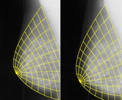
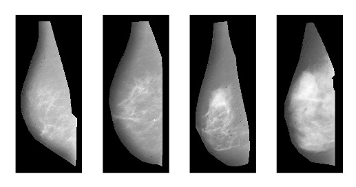
The BreastIQ framework is founded on a segmentation framework the locates the breast region in MLO and CC views. Based on localizing the chest-breast interface and the nipple location, a breast coordinate system is placed on the mammogram1. This is used for defining homologeous points and directions between breasts and for the same breast between repeat mammograms. This is essential for performing focal statistics of mammogram texture and appearance.
Breast density and breast cancer risk
The Gail model is an internationally established tool to establish the risk of breast cancer in clinical practice. Recent reports indicate that the addition of breast density and other risk factors to the Gail model may increase the ability to predict cases of breast cancer.
Among the numerous methodologies for density scoring, the categorical density scoring of the Breast Imaging Report and Data System (BI-RADS©) was originally proposed by the American College of Radiology for quantifying masking effects. Before this, Wolfe Patterns were introduced to assess the risk of breast cancer. Both methods rely on an expert’s categorisation of the mammographic appearance. An interactive threshold measurement methodology that expresses the area of dense tissue as a percentage of the total breast area has advantages in terms of being a continuous score, but still requires the interaction of a radiologist. Recently, qualifying the density as an objective measure based on quantitative calibrated imaging has attracted attention, and it has been shown that quantitative imaging may increase risk segregation.
Biomediq offers stand-alone software in the BreastIQ framework that automatically estimates the breast density from mammograms. It also offers computer support for radiologists scoring of percentage density in a smooth workflow.
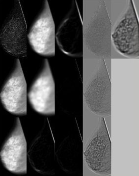
Texture markers MTR and breast cancer risk
Several methodologies have been attempted for quantification of mammographic texture. A very general approach, recording structural components in subjects with known outcome and recognizing these in mammograms to be scored, has shown very promising results2,3,7. Only these BC MTR features, which are part of the Biomediq technology, have the capability to distinguish spatially varying features, and to measure aspects that are not yet understood, but simply observed.
They have demonstrated ability to recognize textural patterns, so that the risk segergation of density is significantly improved. The textural pattern recognition by BC MTR yields and additional odds ratio of 2 from lower to higher quartile.
Texture relating to treatment
The MTR technology may also be used for recognising mammographic texture relating to treatment effects. This has shown incerased mammographic sensitivity compared to density4,6,7. This gives important information relating to the discussion that density increase or change in general may have various structures and show that the textural changes in BC risk lowering and other treatments are alike5.
References
- Brandt, S.S., et al., An anatomically oriented breast coordinate system for mammogram analysis. IEEE transactions on medical imaging, 2011. 30(10): p. 1841-51.
- Nielsen, M., et al., Mammographic Texture Resemblance generalizes as an independent risk factor of breast cancer, in 5th International Workshop on Breast Densitometry and Breast Cancer Risk Assessment, S. Cummings, Editor 2011.
- Nielsen, M., et al., A novel and automatic mammographic texture resemblance marker is an independent risk factor for breast cancer. Cancer Epidemiol, 2010.
- Nielsen, M., et al., Breast density changes associated with postmenopausal hormone therapy: post hoc radiologist- and computer-based analyses. Menopause, 2010. 17(4): p. 772-8.
- Nielsen, M., et al., Low-dose transdermal estradiol induces breast density and heterogeneity changes comparable to those of raloxifene. Menopause, 2009. 16(4): p. 785-91.
- Pettersen, P.C., et al., Parallel assessment of the impact of different hormone replacement therapies on breast density by radiologist- and computer-based analyses of mammograms. Climacteric, 2008. 11(2): p. 135-43.
- Raundahl, J., et al., Quantifying effect-specific mammographic density. Med Image Comput Comput Assist Interv, 2007. 10(Pt 2): p. 580-7.